+91 8878385555
zenithclinics@gmail.com
+91 8878385555
zenithclinics@gmail.com
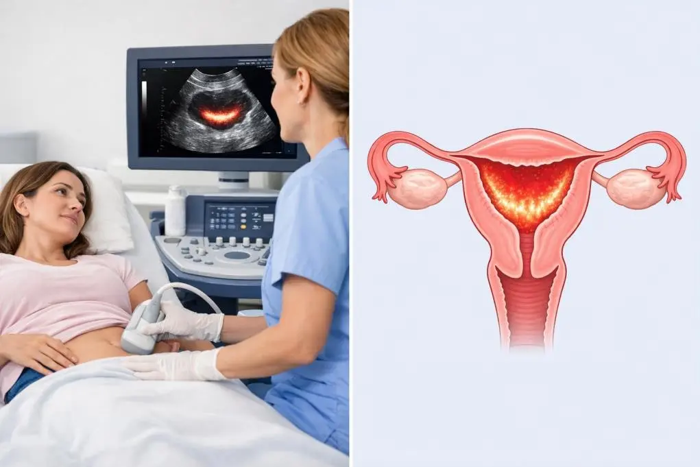
Heavy menstrual bleeding is often ignored as normal, but persistent heavy periods may indicate menorrhagia. This condition can lead to anemia, fatigue, and serious gynecological issues. A pelvic ultrasound helps identify causes like fibroids, thickened endometrium, hormonal imbalance, or early uterine disorders, enabling timely diagnosis and effective treatment.

The thyroid gland plays a crucial role in regulating metabolism, energy levels, heart rate, and overall hormonal balance. Yet, many thyroid problems develop silently and are discovered only when symptoms become noticeable.

What is an ultrasound scan? Ultrasound scanning, or sonography, is a standard procedure in medical practice established more than 30 years ago. It is completely safe and there have never been any known side-effects on the mother or the baby. It functions on the principle of sending out sound waves inaudible to humans via an ultrasound

What is an early pregnancy scan? Early pregnancy scan is one of the most important ultrasound scans in pregnancy because it confirms the pregnancy and verifies the fetus is developing as expected. It can be repeated after a couple of weeks, in order to follow the typical development of your baby and to check if the heartbeat
RPOC (Retained Products of Conception) occurs when placental or fetal tissue remains in the uterus after delivery, miscarriage, or abortion. It can cause persistent bleeding, infection, and long-term complications if not detected early.